
Figure 4.
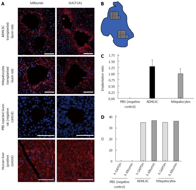
Liver analysis and human cells quantification. A: Immunohistochemistry against human albumin and human uridine diphosphate-glucuronosyltransferase 1A1 (UGT1A1) proteins on serial liver slices in Gunn rats transplanted with adult-derived human liver stem/progenitor cells (ADHLSC) and hHepatocytes. Immunostainings were analyzed by fluorescent microscopy. Slices were fixed and incubated with corresponding primary antibodies. Immunoreactivity was visualized using a 1/500 AlexaFluor594 anti-rabbit antibody solution. Human liver was used as positive control for albumin and UGT1A1 staining whereas negative control consisted of Gunn rat liver injected with PBS. Cell nuclei are stained using DAPI (blue). Bar scale: 200 μm; B: Schematic representation of an immunostained slide. Human albumin positive cells were counted over two randomly chosen areas of the section; C: Mean of human cells repopulation ratio in PBS (negative control), ADHLSC (black column) and hHepatocytes (grey column) transplanted Gunn rats (P > 0.05). Measurement was based on human albumin positive cells counted over two randomly chosen areas of the section; D: Human GAPDH (light grey column) and albumin (dark grey column) detection by RTqPCR in negative control (PBS injected), ADHLSC, and human hepatocyte injected rat livers.