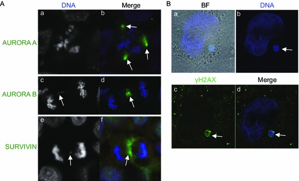
Figure 2.

Aberrant mitosis and micronucleus formation in hPSCs. (A) Immunostaining of euploid HUES1 hESCs for AURORA A (a and b), AURORA B (c and d) and SURVIVIN (e, f). Nuclei are counterstained with DAPI. Panels (a and b) show an example of a multipolar division with multipolar spindles. Note that AURORA A (green staining) is localized to all three spindle poles (white arrows in b). Panels c–f show an example of a chromosomal bridge (white arrows in c and e). Note that AURORA B (d, green staining) and SURVIVIN (f, green staining) are concentrated at the cleavage furrow and the lagging kinetochore (white arrows in d and e). (B) Micronucleus formation (white arrow) in H9 hESCs after inhibition of AURORA B with 50 nm of AZD1152 for 24 h and release from the inhibitor for 24 h. Micronucleus stained positive for γH2AX, a marker of DNA damage (green)
