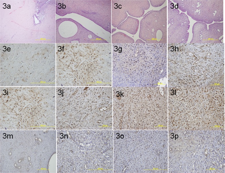
Fig. 3.
a Light microscopic view of right uterine horn in intrauterine adhesion group (group 1). b Light microscopic view of right uterine horn in group 2. c Light microscopic view of right uterine horn in group 3. d Light microscopic view of right uterine horn in group 4. e The immunhistochemical staining of PCNA in group 1. f The immunhistochemical staining of PCNA in group 2. g The immunhistochemical staining of PCNA in group 3. h The immunhistochemical staining of PCNA in group 4. i The immunhistochemical staining of Ki-67 in group 1. j The immunhistochemical staining of Ki-67 in group 2. k The immunhistochemical staining of Ki-67 in group 3. l The immunhistochemical staining of Ki-67 in group 4. m The immunhistochemical staining of VEGF in group 1. n The immunhistochemical staining of VEGF in group 2. o The immunhistochemical staining of VEGF in group 3. p The immunhistochemical staining of VEGF in group 4

