
Fig. 2.
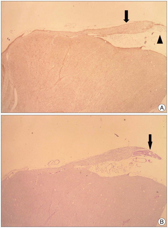
Photomicrograph showing an exiting facial nerve from brain stem. A : Central segment of facial nerve positive to glial fibrillary acid protein (arrow) turned out to be a brown color and peripheral segment is not stained (arrowhead). B : Peripheral segment of facial nerve positive to periodic acid-Schiff (arrow) turned out to be a deep purple color (original magnification, ×10).