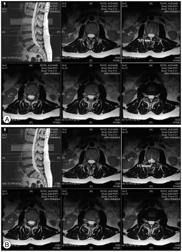
Fig. 3.
The method of measuring zones 4 and 5. A : Zone 4, the distance from the dural sheath to the surface of the ligamentum flavum, is measured in this patient at L1-2 using a T2-weighted axial image. B : Zone 5, the anteroposterior width of the dural sac, is measured in an T2-weighted axial image shown here at L1-2.

