Figure 1.
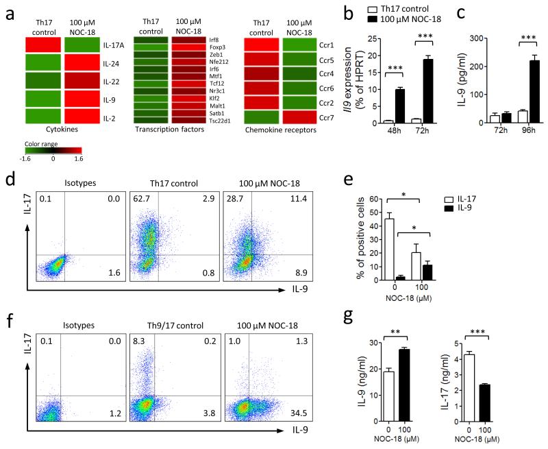
NO inhibits Th17 but enhances Th9 development. Purified BALB/c CD4+ T cells were cultured for 3 d under Th17 polarizing conditions (round-bottom 96-well plate with APC, αCD3, IL-6, TGF-β, IL-23, IL-1β, αIFNγ and αIL-4). NO donor (NOC-18) was added at the beginning of culture. (a) Microarray analysis of differential mRNA expression between cells cultured with or without NO. Full microarray information is provided in Supplementary Fig. 1 and deposited in MIAME (E-MEXP-3959). (b) Il9 mRNA was further quantified by qPCR. (c) IL-9 concentration in the supernatant determined by ELISA. (d) IL-17 and IL-9 producing cells were analyzed by FACS. Numbers in quadrants indicate percentage of cells and are summarized in (e). (f) CD4+ T cells were cultured under Th17/Th9 conditions (αCD3 + IL-4, TGFβ, IL-6, IL-1β, IL-23, anti-IFNγ) ± NO. Percentage of cells producing IL-9 and IL-17 determined by FACS. (g) IL-9 and IL-17 concentrations in the culture supernatant assayed by ELISA. Data are representative of at least 3 independent experiments; Mean ± SEM, n=3; *P<0.05, **P< 0.01, ***P< 0.001 using a 2-way ANOVA (b,c and e) or a 2-tailed unpaired Student’s t test (g).

