Figure 5.
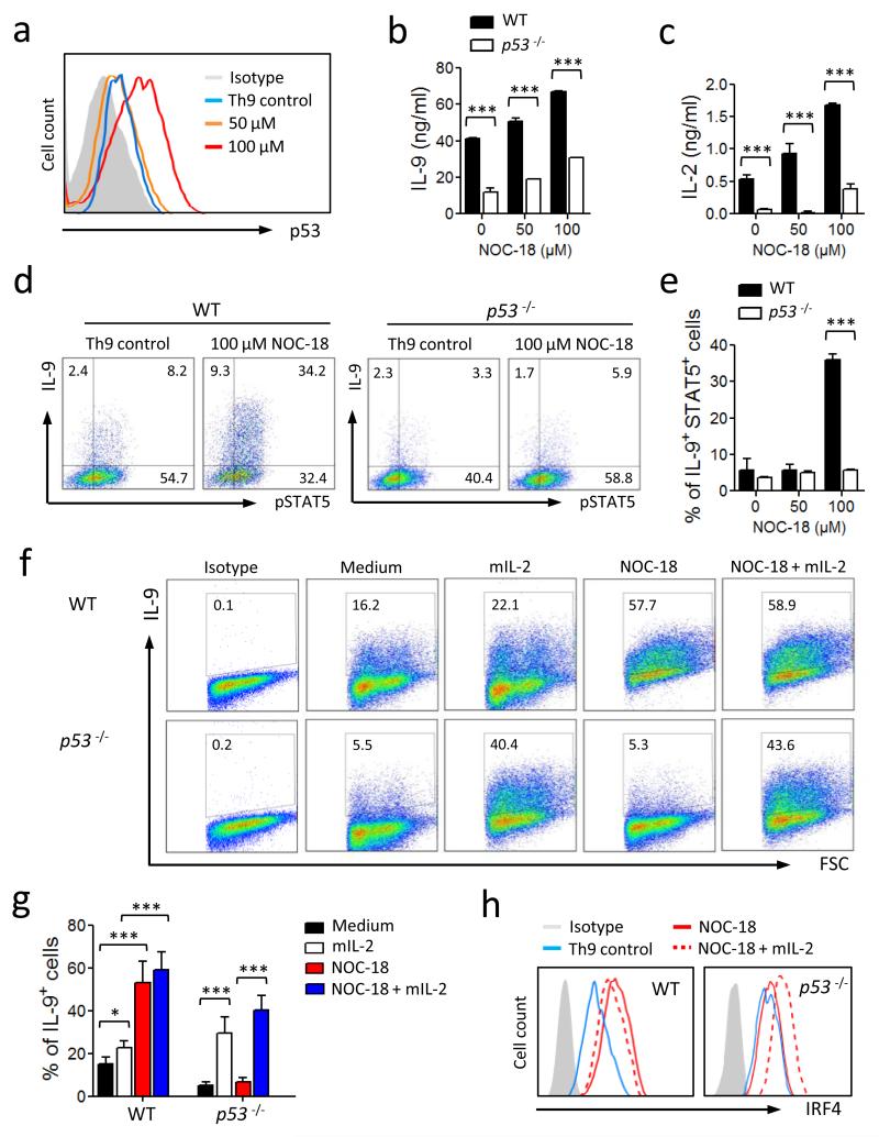
NO-induced up-regulation of Th9 polarization is p53-dependent. CD4+ T cells from WT and p53−/− B6 mice were cultured under Th9 polarizing conditions ± NO. (a) The expression of p53 in WT Th9 cells on day 4 was analyzed by FACS. Concentrations of IL-9 (b) and IL-2 (c) in the culture supernatants of WT and p53−/− cells on day 5 were determined by ELISA. (d, e) Percent of IL-9+ and pSTAT5+ T cells from WT and p53−/− mice polarized under Th9 conditions was analyzed by FACS on day 4. Representative (f) and percent (g) of IL-9+ T cells from WT or p53−/− mice cultured under Th9 conditions in the presence of NO with or without IL-2 are shown. (h) Expression of IRF4 in the Th9 cells from WT and p53−/− mice polarized under Th9 conditions ± NO ± IL-2 was analyzed by FACS. Data are representative or pool of at least 3 experiments, mean ± SEM, n=3, *P<0.05, ***P<0.001 using a 2-way ANOVA.

