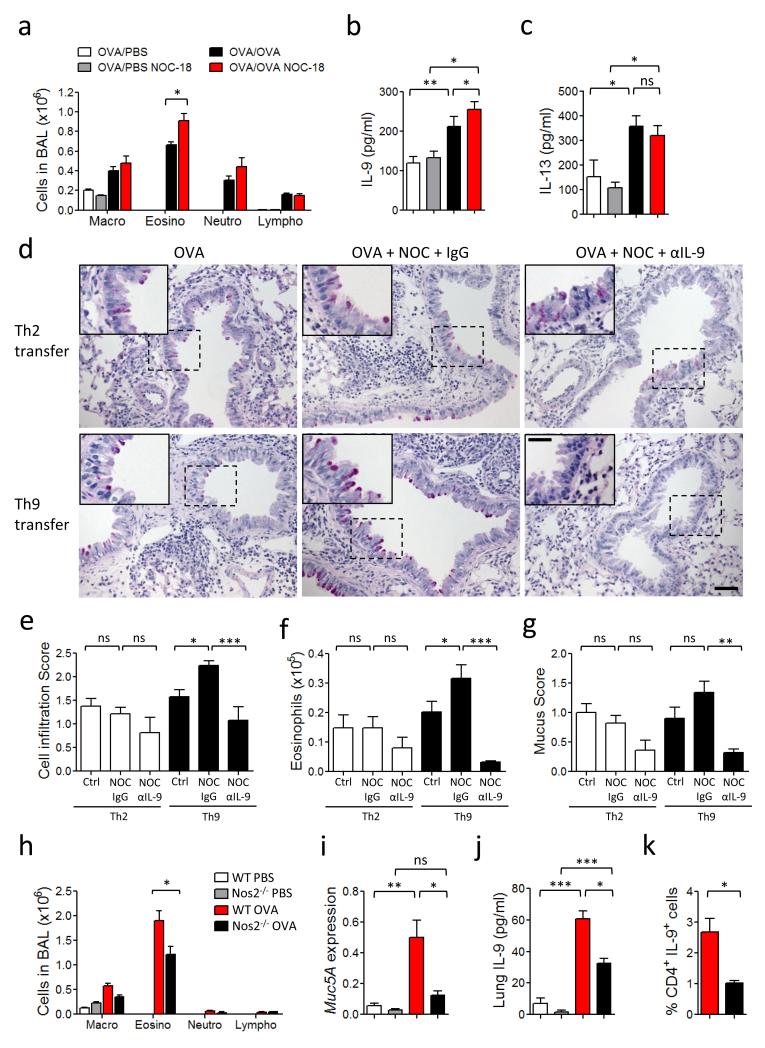
Figure 7.
NO enhances IL-9 production and Th9 frequency and exacerbates allergic airway inflammation in mice. (a-c) OVA-immunized C57BL/6 mice were challenged with OVA and injected s.c. every 2 days with NOC-18. Differential cell counts (a), IL-9 concentration (b) and IL-13 content (c) in the BAL fluid were determined 24 h after the last challenge. (d-g) CD4+ T cells from OT-II mice were polarized to Th9 or Th2 cells and adoptively transferred i.n. to naïve recipient mice which were then challenged i.n. with OVA ± NOC-18. Some mice were injected i.p. with αIL-9 antibody or IgG. PAS staining were performed on lung sections obtained 24 h after the last challenge (d). Scale bars represent 100 μm and 400 μm for the insert. Cell infiltration in the lungs (e), Eosinophils in BAL fluids (f), and mucus score in the lung tissues (g) were measured. (h-k) WT and Nos2−/− mice were immunized s.c. with OVA and challenged i.n. with OVA or PBS. BAL was analyzed by differential cell count (h). Lungs were analyzed for Mucin5A mRNA expression by qPCR (i), and IL-9 concentration by ELISA (j). Percentage of Th9 (CD4+/IL9+) cells in the draining lymph node analyzed by FACS (k). Experiments were performed at least twice; data are mean ± SEM, n=5-6 mice per group; *P<0.05, **P<0.01, ***P<0.001 using 2-way ANOVA.

