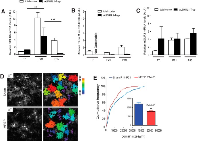
Figure 6.
Developmental growth of cortical astroglial domain is inhibited by in vivo MPEP administration. A–C, Relative ribosome-bound translating mGluR5 (A), mGluR1 (B), and mGluR3 (C) mRNA levels in cortical astroglia compared with corresponding mRNA levels in the total cortex during development. N = 3–4 mice/group. p value was determined in Student's t test. Ribosome-bound translating mRNAs were isolated using TRAP approach. D, Representative images of cortical astroglial domains in EAAT2 tdT reporter mice following MPEP administration. Astroglial domain is color-coded based on the volume size. Scale bar, 40 μm, E, Cumulative frequency curve of cortical astroglial domain size in EAAT2 tdT reporter mice following MPEP administration. The inset bar graph represents the average astroglial domain size. N = 159–173 astroglia/group from multiple mice. p value was determined using the Kolmogorov–Smirnov test.

