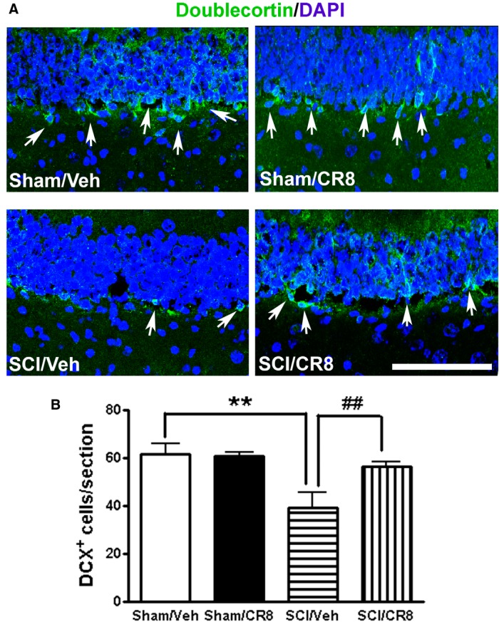
Figure 11.
Distribution and quantification of immature neurons in the adult DG after SCI. A, Representative images showed doublecortin (DCX, green, white arrows) and nuclear staining (DAPI, blue) in the hippocampal DG subregion. Scale bar, 100 μm. B, Quantification of the number of DCX+ cells per DG in a 20 μm section. SCI caused a significant 36% reduction of DCX+ cells at 12 weeks after injury compared with Sham samples. CR8-treated mice showed significantly increased DCX+ cells in the DG. n = 5 images/DG/mouse/6 mice/group. **p < 0.01 versus Sham/Vehicle mice; ##p < 0.01 versus SCI/Vehicle.

