Figure 5.

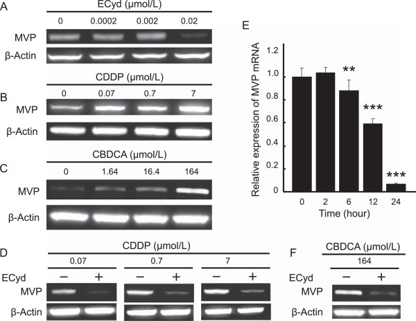
ECyd cancels the induction of MVP protein expression induced by CDDP treatment. A-C) The expression of MVP protein in KB/CDDP(T) cells treated with 0–0.02 μmol/L (IC50 value) of ECyd (A), 0–7.0 μmol/L (IC50 value) of CDDP (B) or 0–164 μmol/L (IC50 value) of CBDCA (C) for 72 hours was analyzed using immunoblot analysis. Equal loading was confirmed by the detection of β-actin. D) The expression of MVP protein in KB/CDDP(T) cells treated with 0–7.0 μmol/L (IC50 value) of CDDP with or without ECyd (0.02 μmol/L) for 72 hours was analyzed using immunoblot analysis. Equal loading was documented by the detection of β-actin. E) KB/CDDP(T) cells were treated with ECyd (0.02 μmol/L) for several terms. The mRNA level of MVP was analyzed using RT-PCR. The Ct value of mRNA was normalized according to that of 18S rRNA as an endogenous control. Columns, mean; bars, SD; **P < 0.01, ***P < 0.001 (n = 3). F) The expression of MVP protein in KB/CDDP(T) cells treated with 164 μmol/L (IC50 value) of CBDCA with or without ECyd (0.02 μmol/L) for 72 hours was analyzed using immunoblot analysis. Equal loading was documented by the detection of β-actin.
