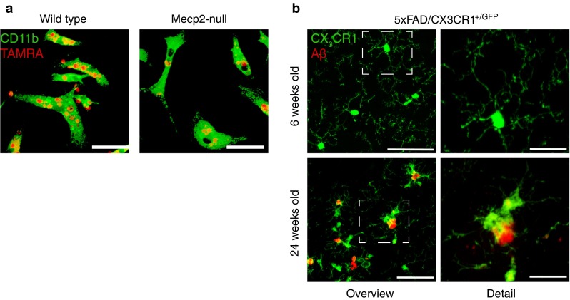
Fig. 1.

Aberrant microglia in models of Rett syndrome and Alzheimer’s disease. a Representative captures of phagocytosing microglia (labeled with anti-CD11b, green) incubated for 2 h with TAMRA-labeled UV-irradiated neural progenitor cells (red). b Representative confocal images of microglia in a pre-depositing 6-week-old 5xFAD transgenic mouse and activated microglia encompassing plaques in a 24-week-old 5xFAD transgenic mouse. Note the shortened and less ramified processes. Green labeling is GFP expressed under the control of the CX3CR1 promoter; red fluorescence depicts Aβ plaques labeled with an anti-Aβ antibody. Scale bars: a 25 µm and b left panel 50 µm; right panel 20 µm
