
Fig. 7.
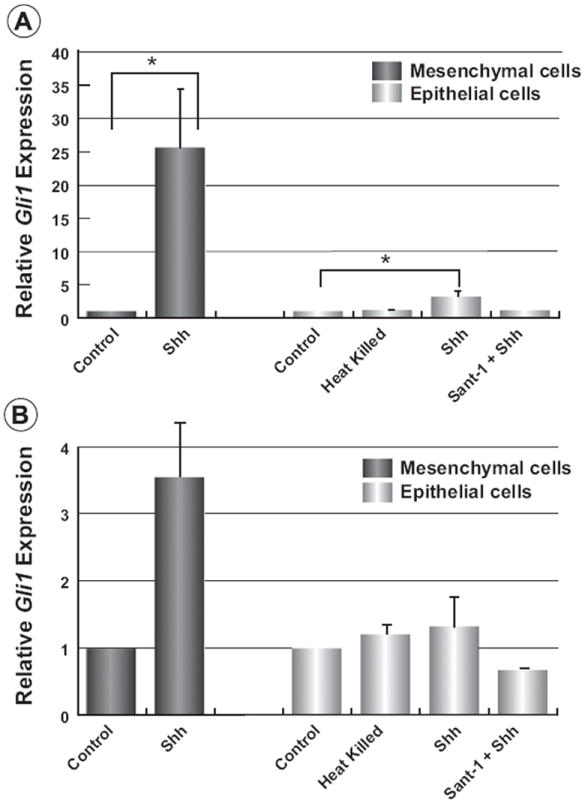
Limited canonical Hh-signalling in primary mammary epithelial cells. Primary mammary epithelial and mesenchymal cells isolated from three monthold wild type mice were serum starved for 24 h and 48 h, respectively. Cells were then stimulated with heat-killed N-Shh peptide, active N-Shh peptide, or N-Shh peptide with 20 nM SANT-1 for 24 h. (A) qPCR reveals that Gli1 is induced 25-fold in mesenchymal by N-Shh. Only a small but significant 3-fold increase in Gli1 was seen in epithelial cells from the same animals, however. (B) N-Shh peptide induced Ptch1 expression 3.5-fold in mesenchymal cells. No induction of mPtch1 was observed for epithelial cells. Data were annalyzed by one-way ANOVA followed by pairwise comparisons of means by Dunnett’s multiple comparisons test. Data are displayed as mean ± SE, *p < 0.05, n = 3.