
Figure 1. Reperfusion significantly increases CaMKII dependent phosphorylation of RyR2 and PLN.
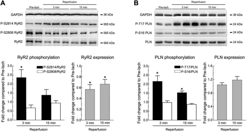
A. Western blots and summary results of CaMKII and PKA-dependent phosphorylation of RyR2 at S2814 and S2808 respectively, and of RyR2 expression, in Langendorff perfused WT mouse hearts subjected to 45 min ischemia followed by various reperfusion times. Comparing with pre-ischemic values (Pre-Isch) there was a significant increase in S2814 phosphorylation at 3 min of reperfusion, without significant changes in the phosphorylation of S2808. A decrease in the expression of RyR2 was present at 3 and 15 min of reperfusion. B. Results of PLN phosphorylation sites during reperfusion. A significant increase in the phosphorylation of the CaMKII-dependent site, T17, was evident at 3 and 15 min of reperfusion with no significant changes in the PKA dependent site, S16. PLN expression did not change relative to pre-ischemic values. GAPDH was used as a loading control. Data represent the average ± SEM of values from 4-9 hearts per group.* P<0.05 vs. the corresponding pre-ischemic values.