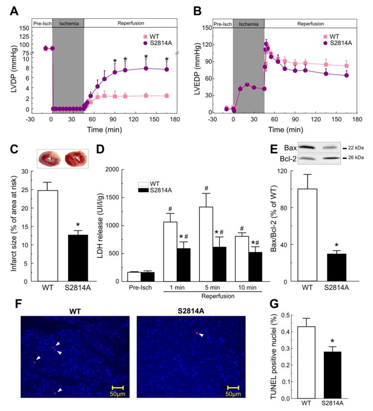
Figure 2. Lack of CaMKII-phosphorylation of RyR2 protects against I/R injury.

Perfused hearts from S2814A mice subjected to I/R (45/120 min), showed a significant improvement of contractile parameters during reperfusion when compared to WT mice, i.e. left ventricular developed pressure (LVDP), was increased (A). Left ventricular end diastolic pressure (LVEDP), showed a trend to be lower in S2814A vs. WT mice, although without reaching significant levels (B). Myocardial viability was enhanced in S2814A vs. WT mice, as reflected by a decrease in the infarct size at the end of reperfusion (C) and in LDH released during the first 10 min of reperfusion (D). Apoptosis was also diminished in S2814A vs. WT mice: Typical immunoblots and overall results after 120 min of reperfusion, depicted in panel (E), showed a decrease in the ratio between the proapoptotic protein Bax and the antiapoptotic protein Bcl2, (Bax/Bcl2). TUNEL assay confirmed apoptotic death decrease in S2814A mice hearts relative to WT. Examples of TUNEL stained cardiac tissue are shown in (F). Total nuclei were stained with DAPI (light blue). Apoptotic nuclei, in red, are marked with arrow heads. Original magnification is 40x. Overall results of this assay are shown in (G). Data represent the average ± SEM of n = 5 - 12 hearts per group.*P<0.05 vs. WT mice. # P<0.05 vs. the corresponding pre-ischemic values.
