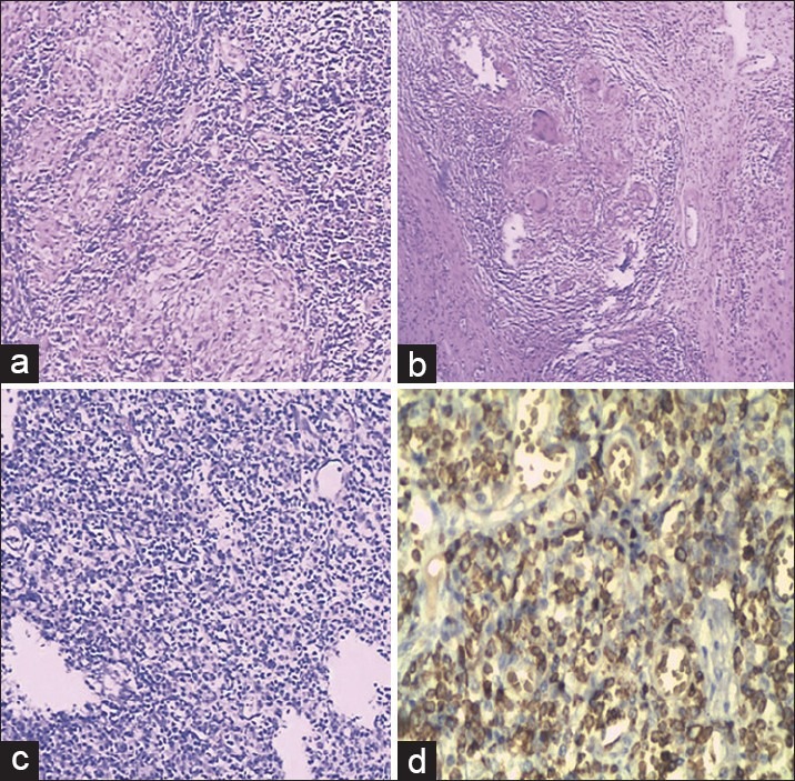
Figure 3.

Pathological features. (a) Caseating granulomas from the mucosa of a patient with ITB [HE, ×100] (b) Large granulomas in the ulcerated mucosa of a patient with CD [HE, ×100]. (c) Atypical lymphoid from the mucosa of a patient with PIL [HE, ×100]. (d) Atypical lymphoid from the mucosa of a patient with PIL [IHC, ×100]
