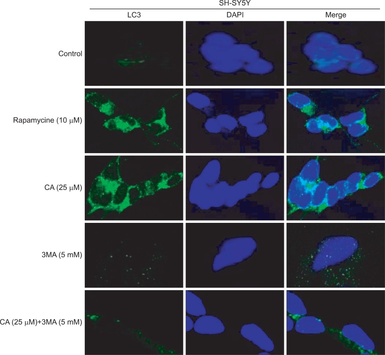
Fig. 4.
CA-induced LC3 expression in autophagosomes. SH-SY5Y cells were treated with 25 μM of CA, 10 μM of rapmycin and 5 mM of 3MA for 24 h and then, were labeled with 40, 6-diamidino-2-phenylindole (DAPI, blue), and Alexa 488 secondary antibody against LC3 (green). LC3 fluorescent puncta were observed in cells by using the confocal microscope.

