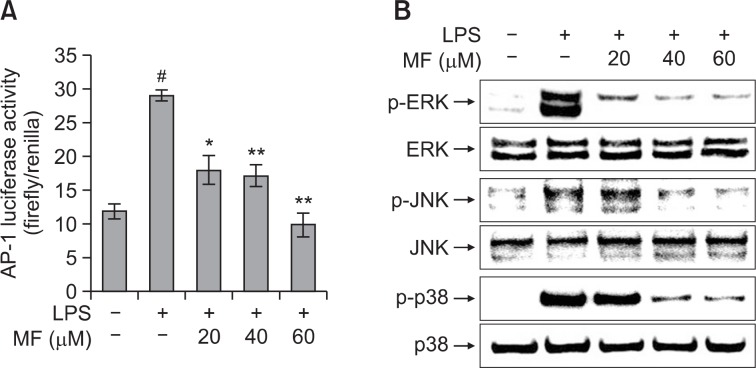
Fig. 4.
Effect of MF on LPS-induced AP-1 activity (A) and phosphorylation of MAPK (B) in RAW264.7 macrophages. (A) After transfection with pAP-1-Luc and pRL-TK, RAW264.7 cells were pretreated with MF (20, 40, and 60 μM) for 1 h and then incubated for 24 h with LPS (1 μg/ml). The AP-1 luciferase activity was determined using a dual-luciferase reporter assay. Error bars represent the mean ± SD. #p<0.001 vs. control, *p<0.01 vs. LPS, **p<0.001 vs. LPS. (B) RAW264.7 cells were pretreated with MF (20, 40, and 60 μM) for 1 h and then incubated for 30 min with LPS (1 μg/ml). The cell extracts were prepared and the phosphorylation of MAPK was determined using western blot analysis.

