Fig. 1.
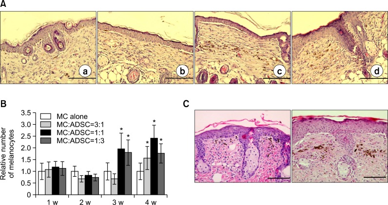
Simultaneous grafting of melanocytes, with or without ADSCs in nude mice. (A) Light microscopy of skin specimens stained with H&E (×400). Skin was grafted with indicated cells, such as melanocytes alone (a, 3.3×10⁴ cells), melanocytes to ADSCs ratio as 3:1 (b), 1:1 (c) and 1:3 (d) for 2 weeks. Scale bar=200 μm (B) Relative number of melanocytes, which was determined by calculating the ratio of melanocyte number cultured in each condition to that cultured melanocyte alone for 1 week, for 4 weeks after grafting of melanocytes, with or without ADSCs as indicated ratios. The number of melanin-containing melanocytes was counted in 5 random microscopic fields (high power, ×400) for statistical analysis. The data represent the mean ± SD of 5 nude mice. *p<0.05 vs. melanocyte monoculture (C) H&E staining of skin specimens 1 week (left) and 2 weeks (right) after grafting of melanocytes alone to compare their residing location. ×400, Scale bar=200 μm.

