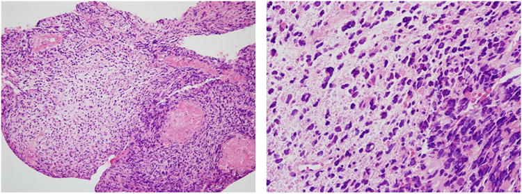
Figure 1.

Biopsy of the nasal mass shows a primitive malignant neoplasm with round and spindled hyperchromatic cells in a loose myxoid matrix, typical of undifferentiated embryonal sarcoma of the liver. The primary liver tumor cells (not shown) showed no expression of epithelial or skeletal muscle markers (pan-cytokeratin, muscle specific actin, desmin). (Hematoxylin and eosin, 25 × (a) and 100× (b) original magnification).
