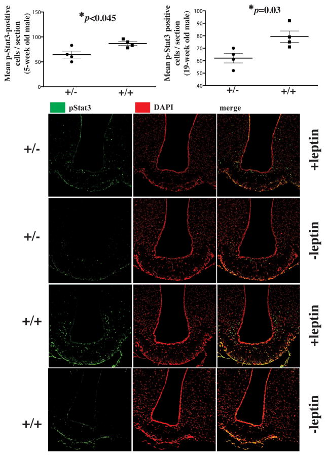
Fig. 3. Reduced leptin signaling in Rpgrip1l+/−mice.
pStat3-positive cells in the hypothalamus of Rpgrip1l+/− and Rpgrip1l+/+ 5-week and 19-week old male mice fed regular chow to whom leptin (2μg/g of body weight) was administered 30 min prior to sacrifice. pStat3-positive cells were counted in 10μm-thick cryosections encompassing the whole arcuate hypothalamus. Nuclear stain is shown in red to improve contrast with pStat3-specific green stain. Error bars represent SEM.

