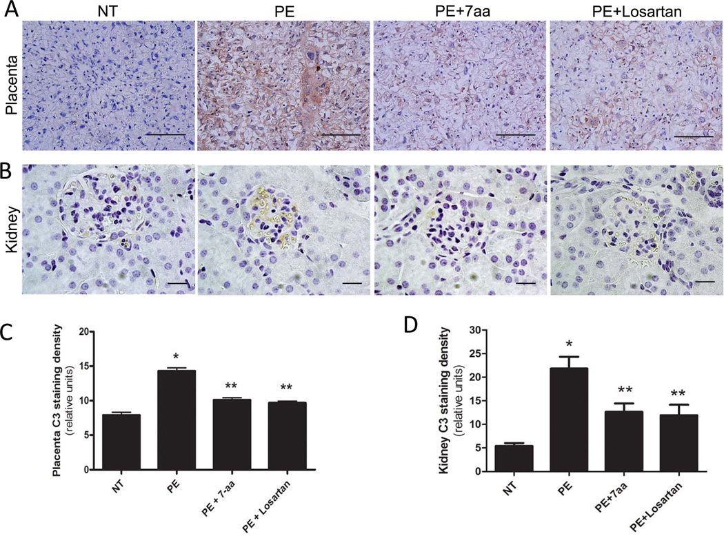
Figure 1. Maternal circulating IgG in preeclamptic women is responsible for increased C3 deposition in placentas and kidneys of PE-IgG-injected pregnant mice via AT1R activation.
Deposition of C3 was examined by immunohistochemistry. C3 expression levels are significantly elevated in (A) placenta (trophoblast cells and endothelial cells; Scale bar = 100µM) and (B) kidney (podocytes of glomeruli; Scale bar = 20µM) of PE-IgG-injected pregnant mice (n=4). Co-injection with losartan (n=9) or the 7-aa epitope peptide (n=9) resulted in substantial reduction of C3 staining. Image quantification shows that C3 staining in both placentas (C) and kidneys (D) is significantly elevated in PE-IgG-injected pregnant mice compared to NT-IgG-injected pregnant mice (n=4). * P < 0.05 compared to NT-IgG injected mice; ** P < 0.05 compared to PE-IgG injected mice.

