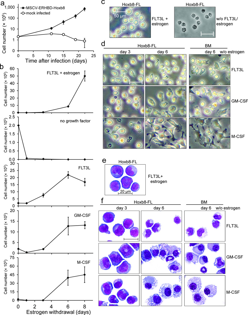
Figure 1. Growth and morphology of Hoxb8–FL cells.
(a) BM cells were infected with an MSCV–based, ERHBD–HOXB8 expressing retrovirus or mock infected and cultured in the presence of estrogen and FLT3L. Cell numbers were determined at time points indicated. Error bars represent standard deviation of cells grown in five individual wells. For viral construct and procedure see also Supplementary Figure 1.
(b) Medium of 2×10⁵ exponentially growing Hoxb8–FL cells was exchanged by medium with indicated factors and cell numbers of live cells were determined at depicted time points. Mean cell numbers obtained after eight days of culture were: FLT3L, 1.6×10⁶; GM–CSF, 1.3×10⁶; M–CSF, 4.5×10⁶; Error bars represent standard deviation of three Hoxb8–FL cell populations.
(c–f) β–estrogen– and FLT3L–containing medium of exponentially growing Hoxb8–FL cells was replaced by medium without growth factor (c), or with FLT3L, GM–CSF or M–CSF, as indicated, and cells were analyzed one day (c) and three and six days (d,f) later by phase contrast microscopy in cell culture (c, d) or after cytospin and May–Grünwald/ Giemsa staining by bright field microscopy (e, f). Unfractionated BM cells were cultured in parallel for six days under the same conditions as described for Hoxb8–FL cells and are shown for comparison. Scale bars: (c, d) = 50 µm, (e, f) = 20 µm.

