Figure 5.
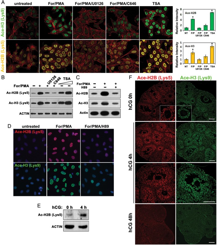
Pre-ovulatory LH surge induces histone acetylation in granulosa cells through ERK1/2 and CBP activity. (A–D) Immunofluorescence (A and D) and western blotting (B and C) results for acetylated histone H2B (Lys5) and H3 (Lys9) levels in cultured GCs treated with the indicated reagents. The concentrations used were: forskolin, 10 µM; PMA, 20 nM; U0126, 10 µM; TSA, 2 µM; and H89, 10 µM. Immunofluorescent signal intensities were quantified using Image J software. *P < 0.001 when compared with sample 1 from left. (E) Western blotting results for acetylated histone H2B (Lys5) levels in mouse ovaries before and after hCG treatment. (F) Immunofluorescence results for acetylated histone H2B (Lys5) and H3 (Lys9) levels in mouse ovaries before and after hCG treatment. Scale bar = 100 µM.

