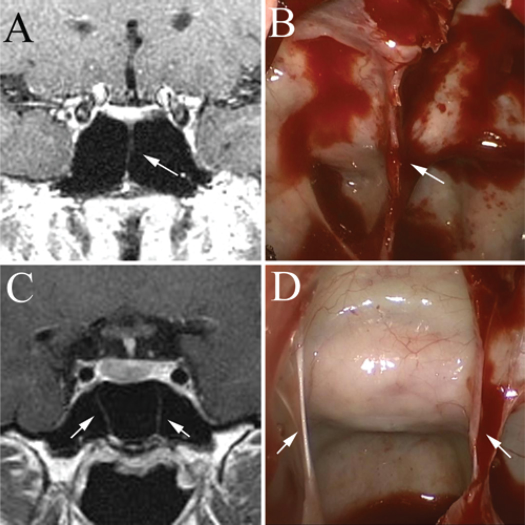
Fig. 10.
Examples of intraoperative findings–imaging correlation of vertical septa in patients with simple sphenoid sinus morphology. The presence of 1 midline vertical septum (A and B, white arrow) can be used to identify the anatomical midline. The presence of 2 symmetric vertical septa (C and D, white arrows) can typically be used to identify the lateral boundaries of the sellar exposure and extrapolate the midline location.

