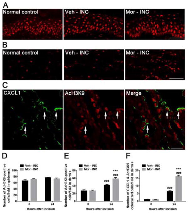
Figure 4.
Levels of acetylated histone H3 at lysine 9 (AcH3K9) in skin tissue at 1 day after incision. (A) Levels of acetylated H3K9 were unchanged in the epidermal layer after incision in both vehicle and morphine pretreated groups. Scale bar: 50 μm. (B) Expression of AcH3K9 was increased after incision and further increased in mice treated with morphine in the dermal layer. Scale bar: 50 μm. (C) Double immunostaining showed CXCL1 (green) and AcH3K9 (red) were colocolized (arrows) in the dermal layer at 1 day after incision in mice treated with morphine. Scale bar: 20 μm. (D) Quantification of AcH3K9 positive cells in the epidermis. (E) Quantification of AcH3K9 positive cells in the dermis. (D) Quantification of both CXCL1 and AcH3K9 positive cells in the dermis. Values are displayed as the mean ± SEM, n = 4, ### p<0.001 vs. day 0 (before incision); *** p<0.001 vs. vehicle treated group. Veh=vehicle; Mor=morphine; INC= incision.

