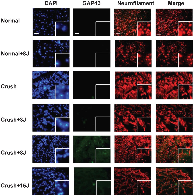
Figure 6. Detection of GAP43 expression in the sciatic nerve of 808-nm LLLT laser-treated distal site using immunofluorescent staining.
Groups: sham-operated rats without (normal) or with 8 J/cm2 LLLT (normal+8J) and rats with sciatic nerve crush injury without (crush) or with LLLT at 3 J/cm2 (crush+3J), 8 J/cm2 (crush+8J) or 15 J/cm2 (crush+15J). Sections were labeled with DAPI (blue), GAP43 (green) and neurofilament (red), which is specifically expressed in neurites. Original magnification: 100×. White boxes show the enlarged views with a magnification of 400×. Scale bar, 200 um.

