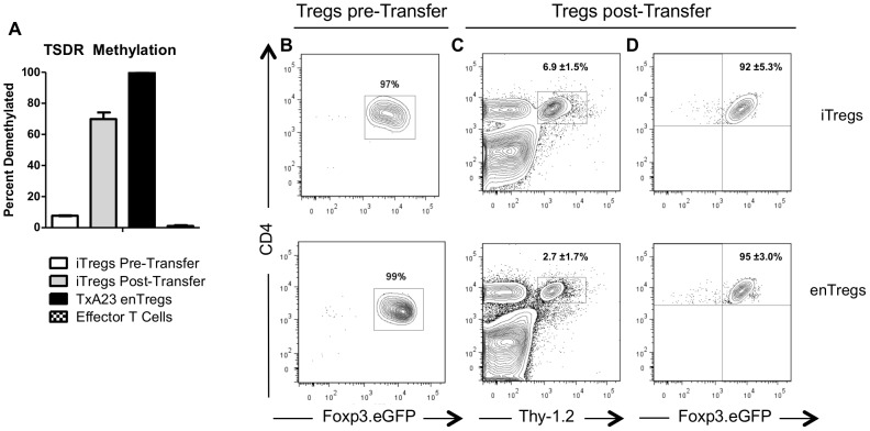
Figure 1. iTregs demethylated the Treg specific demethylated (TSDR) region and maintained Foxp3 expression.
(A) The methylation status of the Foxp3 TSDR of iTregs prior to and after transferring into TxA23 mice. Also shown are enTregs (CD4+Foxp3+) and conventional (CD4+Foxp3-) cells from TxA23 mice. Flow cytometric analysis of (B) the purity of enTregs and iTregs prior to transferring into TxA23 mice, (C) the percentage of Thy1.2+ transferred Tregs in the gastric mucosa, and (D) the expression of Foxp3.eGFP by the Thy1.2+ Tregs one week after transfer. Data shown represents 2 mice for the enTreg group and 3 mice for the iTreg group from two independent experiments.

