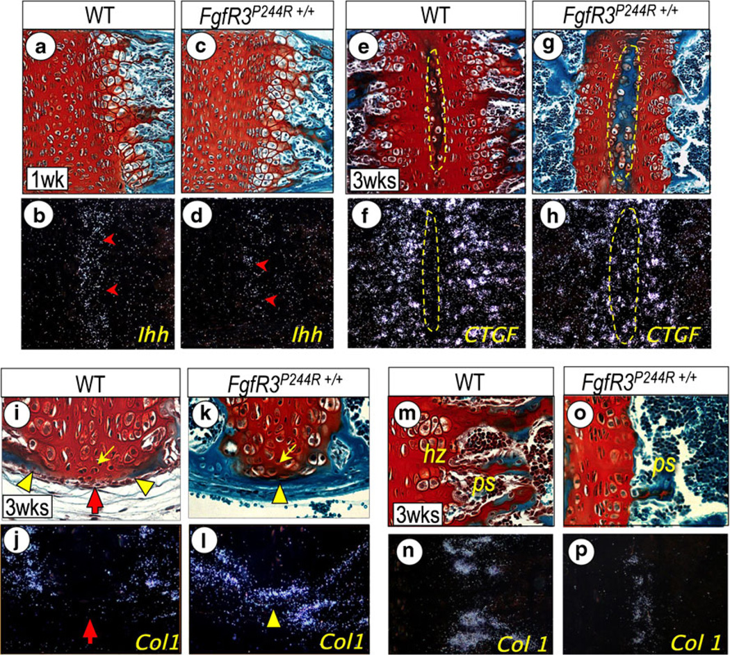
Fig. 5.
Endochondral ossification (a–d), secondary ossification center development (e–h), perichondrial ossification (i–l), and primary spongiosa bone formation (m–p) are shown in wild-type FgfR3P244R+/+ mutant basicranial synchondroses. Parasagittal serial sections of intersphenoidal synchondrosis (ISS) from 3-week-old wild-type (i, j) and mutant (k, l), and spheno-occipital synchondroses (SOS) from 1-week-(a–d) and 3-week-old (e–n) wild-type (a, b, e, f, m, n) and mutant (c, d, g, h, o, p) mice were processed for staining with fast green/Safranin O (a, c, e, g, i, k, m) and in situ hybridization with the indicated genes (b, d, f, h, j, l, n). Notice the marked reduction of Ihh expression in the prehypertrophic zone (d, arrowhead) in mutant SOS. Note also that, in 3-week-old mutant SOS, the shortened growth plate is accompanied with the accelerated development of the secondary ossification center (e, g); indicated with a dotted yellow circle), which is recognizable by the activation of CTGF (h). Precocious perichondrial ossification is visible in the ISS from 3-week-old mutant as shown by fast green staining (k) and in situ hybridization with Type I collagen (Col 1; l) along the entire length of the synchondrosis cartilage. In wild-type ISS, the perichondrium flanking resting chondrocytes (yellow arrow) remains unossified (red arrow) at this time, as indicated by the lack of Col1 and other osteogenic gene (data not shown) expression (i, j). Notice that the chondrocytes flanking the perichondrium in mutant ISS have the appearance of prehypertrophic chondrocytes (k, yellow arrow). Notice the significant reduction of hypertrophic chondrocyte zone (hz) and primary spongiosa (ps) at the chondro-osseous border in 3-week-old mutant SOS (o) compared with wild-type SOS (l), which is identifiable by the significant reduction of Col 1 (m, p). Other genes required for chondro-osseous transition are also drastically reduced (data not shown). (Adopted from Developmental Dynamics 240 (11):2584–2594, 2011)

