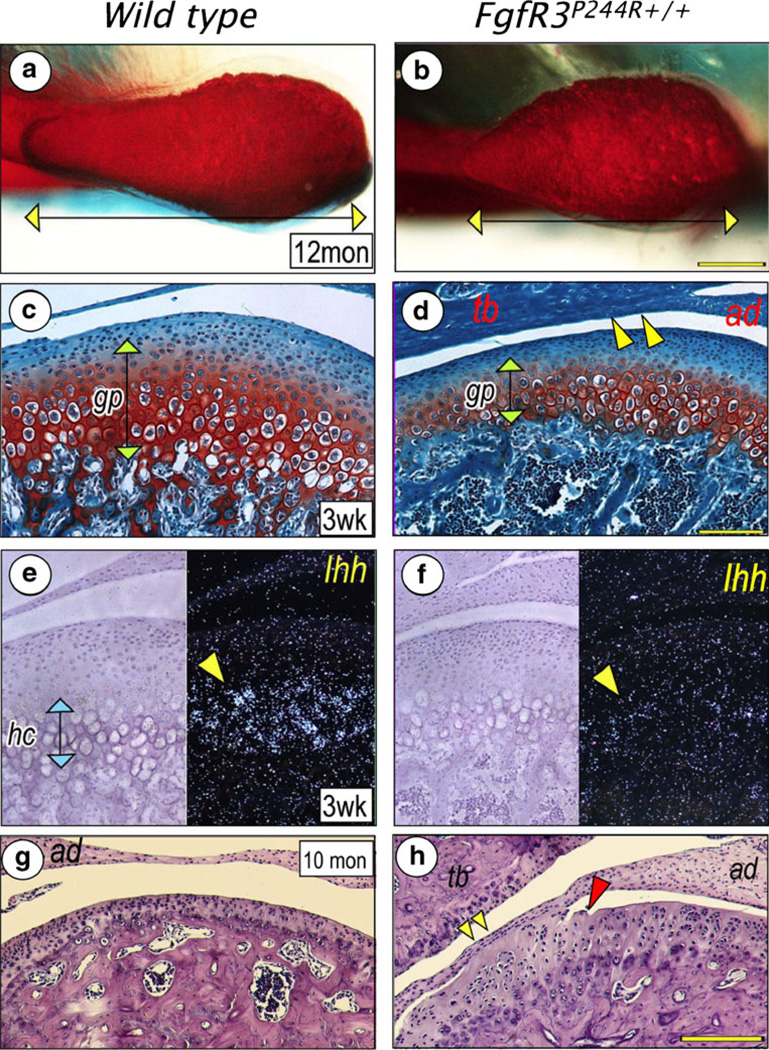
Fig. 6.
Defective development and arthritic changes of the temporomandibular joint in FgfR3P244R mutant mice. A superior view of the condylar head shows that the mesial half is nearly absent in the mutant mice (b) compared with a wild type (a). The mutant mice also show significantly reduced growth plate-like cartilage (gp) of the mandibular condyle (d) and Ihh expression in hypertrophying chondrocytes (hc) (f), compared to a wild type (c, e). Articular discs (ad) of the mutant mice adhere to articular eminence of the temporal bone (tb) (d, yellow arrowheads) or condylar surface (h, yellow arrowheads). Also, the articular surface of the mutant condyle presents fissure formation (h), red arrowhead). Neither disc adhesion nor fissure formation are observed in wild type (c,g). (Adopted from J Dent Res, 2012)

