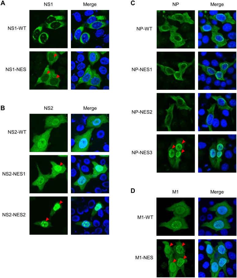
Figure 2. Intracellular localization of NES mutant proteins.
HeLa cells were grown on cover glass and transfected with wild-type NS1 or NS1-NES mutant plasmid (A), wild-type NS2 or NS2-NES mutant (NS2-NES1 or NS2-NES2) plasmid (B), wild-type NP or NP-NES mutant (NP-NES1, NP-NES2, or NP-NES3) plasmid (C), or wild-type M1 or M1-NES mutant plasmid (D) for 48 h before immunofluorescence staining with anti-NS1 MAb, anti-NS2 Ab, anti-NP MAb, or anti-M1 MAb (respectively) followed by an Alexa Fluor 488-conjugated secondary antibody and Hoechst 333342. The cells were then observed under a confocal laser-scanning microscope. The red arrow heads indicate the localization change of NES mutant proteins (compare with wild-type proteins).

