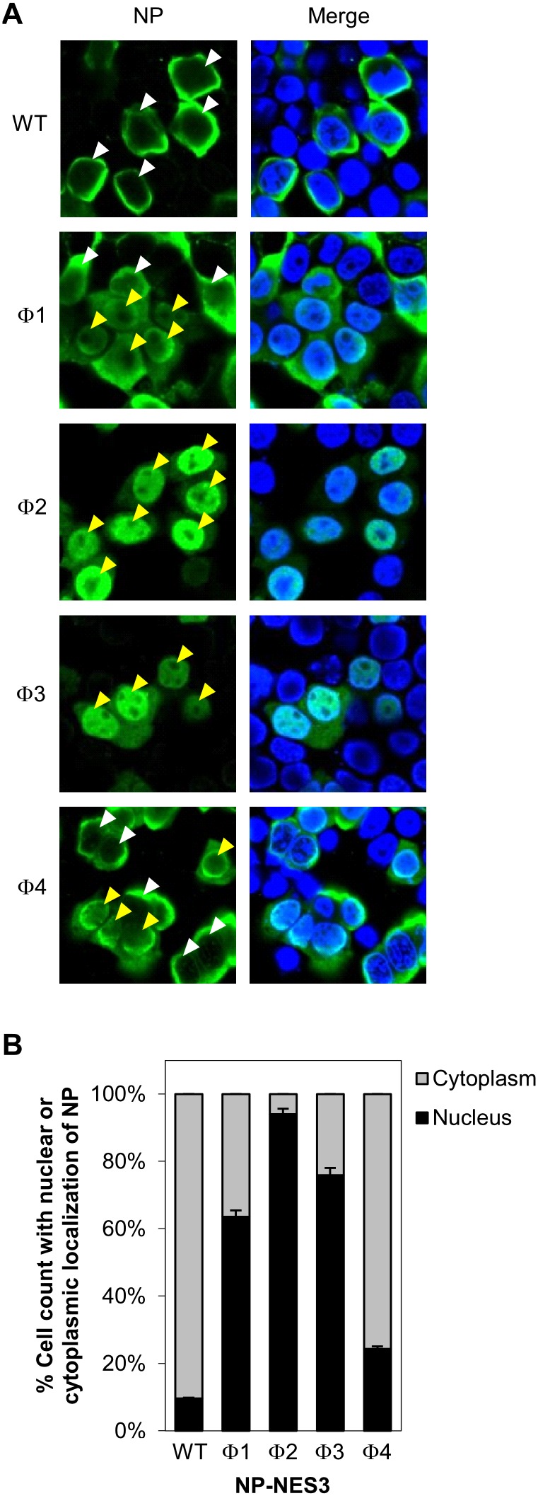
Figure 5. Intracellular localization of NP-NES3 mutant proteins.
(A) HeLa cells were grown on cover glass and transfected with pCAGGS encoding wild-type NP-NES3 or its mutants (Φ1, Φ2, Φ3, or Φ4) for 48 h before immunofluorescence staining with an anti-NP MAb followed by anti-mouse Alexa Fluor 488 and Hoechst 333342. The cells were then observed under a confocal laser-scanning microscope. The white and yellow arrow heads indicate predominant localization of NP in the cytoplasm (cytoplasmic staining > nuclear staining) and nucleus (nuclear staining > cytoplasmic staining), respectively. (B) Nuclear localization of NP wild-type and NP-NES3 mutants from A. Data are presented as the percentage (± SD) of total cell count with predominant nuclear or cytoplasmic staining of NP from five separate fields.

