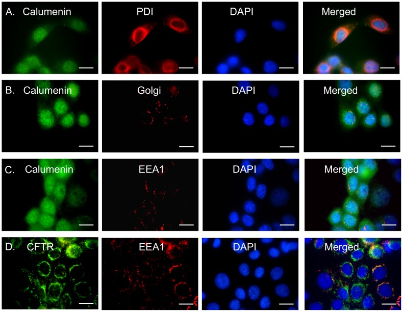
Figure 8. Calumenin trafficking is altered in CFBE41o- cells expressing F508del-CFTR.
Calumenin or CFTR (green) in CFBE41o- wild-type or F508del cells were visualized using fluorescence microscopy. Second labeling was performed for either PDI (an ER marker). Golgi marker or EEA1 (marker for endosomal vesicles) (red). The nuclei were stained blue with DAPI. Colocalisation of green and red pixels was detected in merged images (yellow). A. Calumenin and PDI (for ER staining) B. Calumenin and Golgi and C. Calumenin and EEA1 in CFBE41o- cells expressing F508del-CFTR. D. CFTR and EEA1 in CFBE41o- cells expressing F508del-CFTR. Scale bar: 20 µm.

