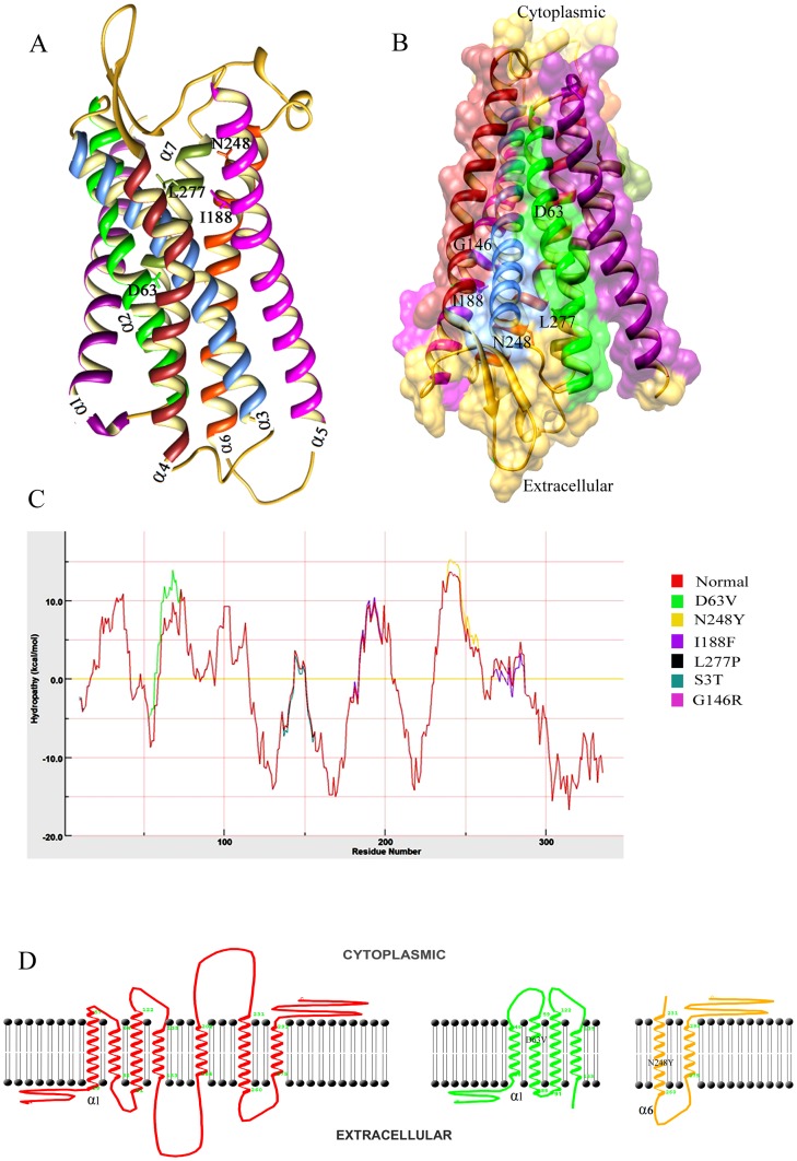
Figure 2. Characterization of LPAR6 specific mutations at structure level.
(A) 3D representation of LPAR6structure in ribbon form. Individual α-helices are indicated by distinct colors: α1, magenta; α2, green; α3, cornflower blue; α4, brown; α5, pink; α6, orange red and α7, olive. β-sheets are indicated by yellow color. (B) Schematic representation of LPAR6 secondary structures indicating the directionality of β-sheets to the extracellular part, while narrow end of α-helices points to the cytosol, making a groove-like structure. Corresponding positions of known amino acids undergoing substitutions are indicated which show that I188, N248 and L277 lie to the extracellular part of α-helices, while D63 and G146 residues are near the cytosolic part. Surface view of groove-like structure is shown to visualize the residual positions. (C) Hydropathy plot analysis of normal and mutated LPAR6 amino acids performed by MPEx tool. (D) Membrane spanning profile studies of individual α-helices for LPAR6WT, LPAR6D63V and LPAR6N248Y.

