Figure 6.
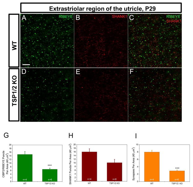
Sensory epithelium of utricle stained with synaptic markers in WT and TSP1/2 mutants. Counts were statistically analyzed with a one-way ANOVA followed by Scheffe’s post hoc test. A – C, WT utricle stained with RIBEYE (A) and postsynaptic SHANK1 (B) at P29. Overlay view is shown in C. D – F, TSP1/2 double mutant utricle stained with presynaptic ribbon marker RIBEYE (D) and postsynaptic SHANK1 (E). Colocalization of both markers is shown in F. G, Synaptic ribbon counts per 40 μm2 area of utricle in TSP1/2 KOs and WT animals. Ribbon numbers in TSP1/2 mutants were lower than WT (P = 0.001). H, Quantification of the postsynaptic marker SHANK1. No significant difference was observed between WT and TSP1/2 mutants. I, Average number of synapses determined by presynaptic RIBEYE that colocalized with postsynaptic SHANK1 per 40 μm2 in WT and TSP1/2 mutant mice. Number of synapses was reduced in TSP1/2 mutants compared to WT (P = 0.000). Number of animals tested (n) per genotype and type of staining are indicated on the graphs. Scale bar in A is 10 μm and applies to B – F. Results are expressed as mean ± SEM. Significant differences are indicated by * for P < 0.05, ** for P < 0.01 or *** for P < 0.001.

