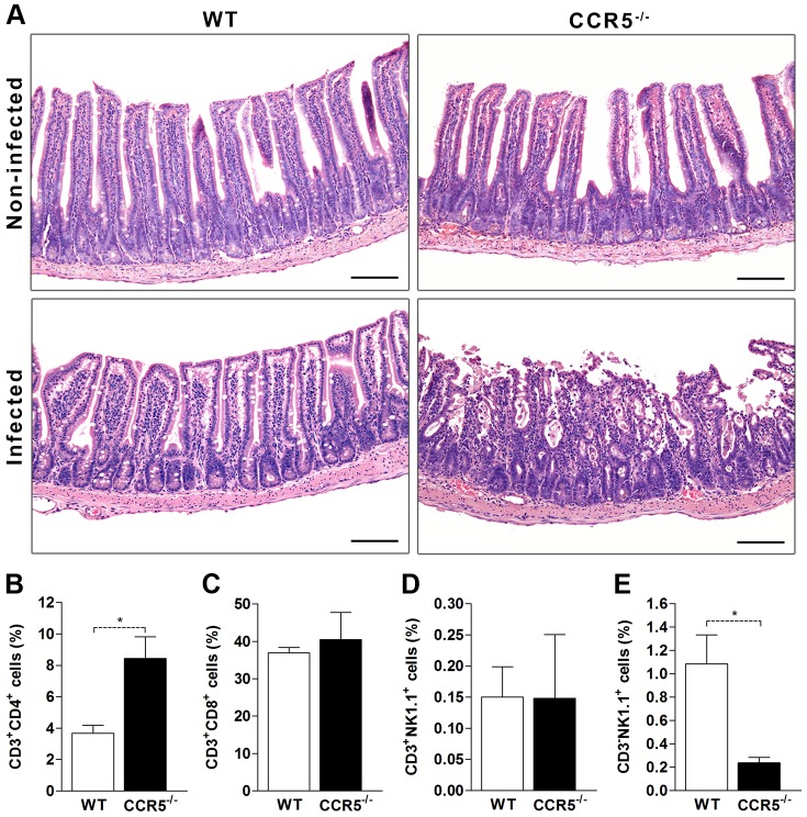
Figure 5. Toxoplasma gondii leads to inflammation, cellular infiltrate and ileum necrosis in the absence of CCR5.
WT and CCR5-/- mice were infected with 5 cysts of T. gondii and at day 8 pi, small intestine was harvested, formalin-fixed, paraffin-embedded, stained with Hematoxylin and Eosin (H&E) and analyzed by light microscopy (A). The leukocytes were isolated from lamina propria of small intestine of WT and CCR5-/- mice at day 8 after T. gondii infection and characterized by flow cytometry. The percentage of CD3+CD4+ T (B), CD3+CD8+ T (C), CD3+NK1.1+ (D) and CD3-NK1.1+ (E) cells was evaluated by FlowJo software. Data represent the mean ± SEM of results from three to five mice per group and are representative of two independent experiments. NI: non-infected. * p<0.05 compared to WT mice.

