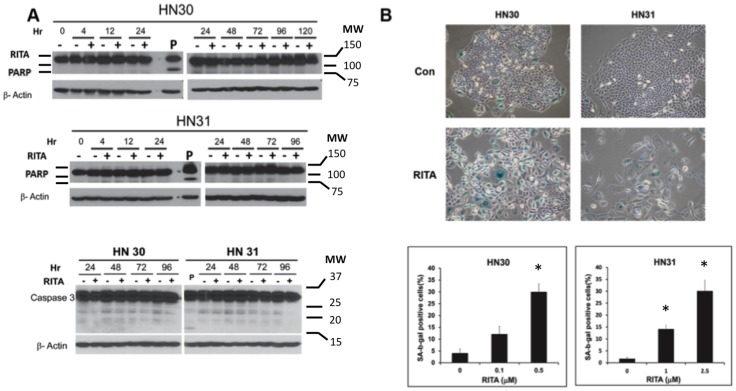
Figure 2. Effects of RITA on proliferation of head and neck cancer cells.
(A) PARP and caspase 3 cleavage were examined via western blotting after exposure of cells to RITA at 1 µM for the indicated periods. P, staurosporine (1 µM for 8 hours) was used as a positive control. (B) HN30 and HN31 cells seeded in 6-well tissue culture plates were treated with the indicated concentrations of RITA for 5 days, after which cells were fixed and stained for senescence-associated -β-galactosidase (SA-β-gal) activity. Representative images from three experiments with similar results are shown. In each treated or untreated well, five random field selections were made and the number of cells with senescent morphology and with blue staining were counted under 20X magnification (Olympus IX71). * - indicates p<0.05 versus untreated control.

