Figure 1.

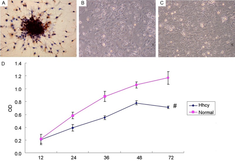
HCY suppresses the activity of cultured hippocampal neurons. A: The results of NSE immunohistochemical staining, in which the NSE immunoreactions complexes were shown as brown and in granulate. The positive NSE granulates were distributed in the cytoplasm and major nerve process, with the glia unstained. B: 1×10⁵ neurons seeded into 6-well plates, incubated with HHcy (1000 μM) for 72 h and then photographed by inverted phase contrast microscope. C: Neurons incubated without HHcy, photographed by inverted phase contrast microscope. Compared with B1, the amount of neurons in B2 was obviously larger. D: The results of MTT Assay for the activity of neurons with or without Hhcy (normal group and Hhcy group) in 12, 24, 36, 48 and 72 hours. The activity of normal neurons in 72 h was higher than that of Hhcy group. #P < 0.05 VS normal group.
