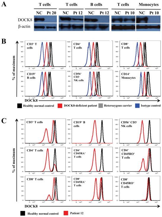
FIG 1.
DOCK8 immunodeficient patients having T cell reversions. A, Immunoblotting for DOCK8 or β-actin proteins in primary T cells, B cells, or monocytes. NC, normal healthy control. Patient 20 has a homozygous large deletion. Patients 12 and 10 have somatic repair. B, Representative flow cytometry histograms showing intracellular DOCK8 expression in gated subsets from a normal healthy control (black), DOCK8 heterozygous carrier (grey), Patient 19 with a large homozygous deletion (red), or isotype control staining (blue). C, Histograms are from Patient 12 who has somatic repair.

