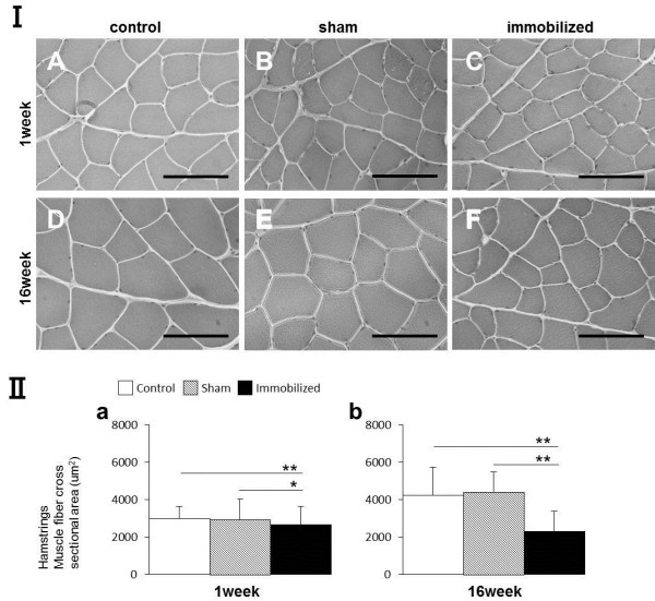
Figure 4.

Morphological analysis and myofiber CSAs measurements (μm2) of the hamstring muscle. (I) The CSA of the hamstring muscles of control, sham-operated, and immobilized knees after 1 and 16 weeks. (A, D) Control group. (B, E) Sham-operated knees. (C, F) Immobilized knees. Scale bars represent 100 μm (microscope magnification: ×400). (II-a, b) Measurement of the CSA (μm2) of the hamstring muscles (only one displayed). Values are presented as mean ± SD; *P < 0.05; **P < 0.001. The CSA in the immobilized knees was lower than that in the control or sham knees at both time points.
