
Figure 7.
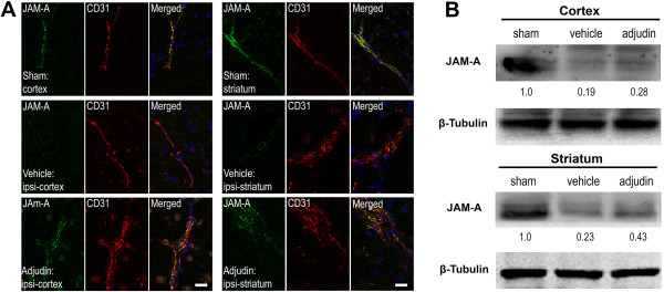
Adjudin inhibited ischemia/reperfusion induced blood-brain barrier destruction assayed by tight junction protein JAM-A. (A) Immunofluorescence staining for JAM-A (green) and CD31 (red) in the cerebral cortex and striatum from mice that underwent sham surgery (sham, top panel), transient middle cerebral artery occlusion (tMCAO) followed by vehicle treatment (vehicle, middle panel) and tMCAO followed by adjudin treatment (adjudin, bottom panel) 3 d after reperfusion. Merged images of JAM-A and CD31 staining were also shown. Scale bar = 100 μm. (B) Representative western blot for JAM-A levels in the cerebral cortex and striatum from mice of the sham, vehicle and adjudin groups. Densitometric value of the protein bands normalized to the respective β-tubulin was also shown.