Fig. 5.

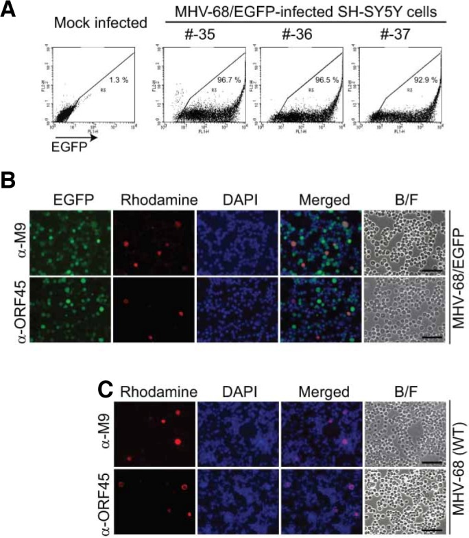
Both productive and latent replications of MHV-68/EGFP in prolonged culture of infected SH-SY5Y cells. (A) Persistent EGFP expression in the majority of MHV-68/EGFP-infected SH-SY5Y cells. MHV-68/EGFP-infected SH-SY5Y cells were grown for 35–37 passages from the initial infection and assayed by flow cytometry for EGFP expression. Mock-infected SH-SY5Y cells were used as the negative control. (B, C) Sporadic expression of lytic viral proteins in the infected SH-SY5Y cells. MHV-68/EGFP- or WT-infected SH-SY5Y cells were grown for 34 or 20 passages from the initial infection, respectively. Immunofluorescence assays were perfor-med using antibodies against ORF45 and M9 viral lytic proteins in the infected cells under a fluorescence microscope. The representative images are shown. B/F indicates the images from the bright field.
