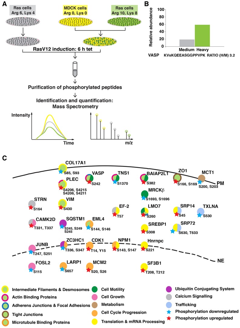
Fig. 1.
Experimental outline of the SILAC screening. (A) MDCK pTR-GFP-RasV12 cells were labeled with medium (Arg 6, Lys 4) or heavy (Arg 10, Lys 8) arginine and lysine, and normal MDCK cells were labeled with light (Arg 0, Lys 0) arginine and lysine. Cells were plated either as Ras cells alone or as a 1∶1 mixed culture of heavy-labeled Ras∶MDCK cells. After 2 h, cells were incubated with tetracycline for 6 h to induce RasV12 expression. Phosphopeptides were isolated and analyzed by mass spectrometry. (B) Relative abundance of the VASP peptide from each experimental condition. The small ‘s’ represents the detected phosphorylation site. (C) Overview of proteins in which phosphorylation was identified as being upregulated (red star) or downregulated (blue star) in Ras cells upon co-incubation with normal cells. The key known functions of the selected proteins based on Gene NCBI, UniProt and PhosphoSitePlus databases are color-coded. Abbreviations of protein names are in agreement with those listed in the Gene NCBI database. PM, plasma membrane; NE, nuclear envelope.

