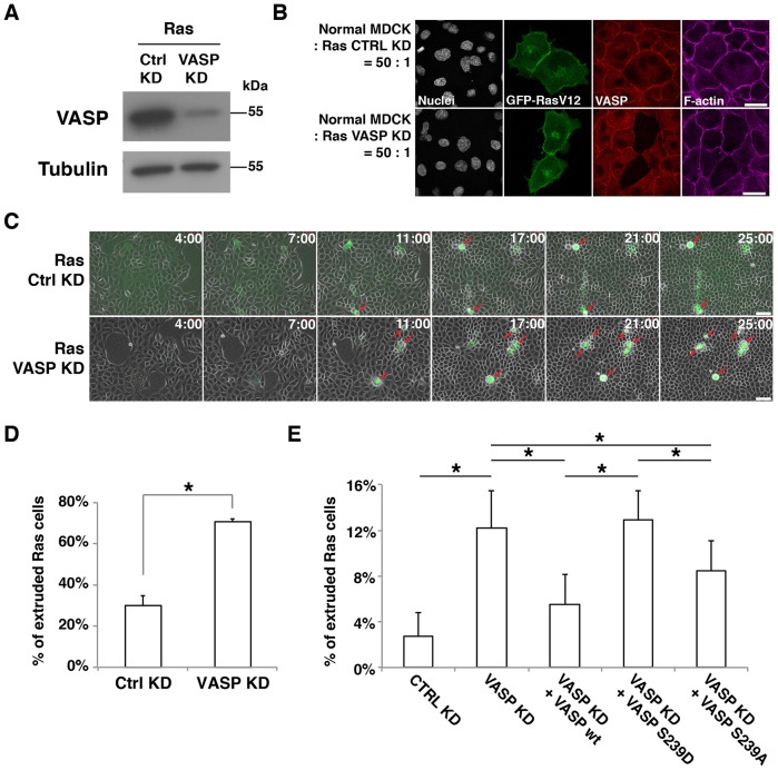
Fig. 3.
Phosphorylation of VASP at S239 promotes apical extrusion. (A) Knockdown of VASP in MDCK pTR-GFP-RasV12 cells. Ras cells were transfected with control siRNA (Ctrl KD) or VASP siRNA (VASP KD). Cell lysates were examined by western blotting with the indicated antibodies. (B) Immunofluorescence of VASP-knockdown Ras cells. Ras cells transfected with control siRNA or VASP siRNA were co-cultured with normal cells on glass. After induction of GFP–RasV12 expression with tetracycline for 8 h, cells were stained with anti-VASP antibody (red) and Alexa-Fluor-647–phalloidin (purple). Scale bars: 25 µm. (C) Time-lapse observation of apical extrusion of Ras cells. Ras cells were transfected with control siRNA or VASP siRNA and co-cultured with normal cells on collagen gels. Images were extracted from the representative time-lapse movies, and the indicated time reflects the duration of the tetracycline treatment. Red arrows indicate extruded Ras cells. Scale bars: 50 µm. (D) Quantification of the apical extrusion of Ras cells from time-lapse analyses (25 h). Data show the mean±s.d. (two independent experiments; n = 174 ctrl KD cells, n = 163 VASP KD cells). (E) The effect of expression of VASP proteins in VASP-knockdown RasV12-transformed cells on apical extrusion. Ras cells were transfected with siRNAs, followed by transfection with the wild-type (wt) VASP, VASP S239D or VASP S239A construct. Note that exogenously expressed human VASP is resistant to the siRNA that targets canine VASP. The transfected Ras cells were co-cultured with normal cells, followed by tetracycline treatment for 15 h. Data show the mean±s.d. (four independent experiments; n = 390 Ctrl KD cells, n = 388 VASP KD cells, n = 373 VASP KD+VASP WT cells, n = 343 VASP KD+VASP S239D cells, n = 324 VASP KD+VASP S239A cells); *P<0.05.

