Fig. 5.
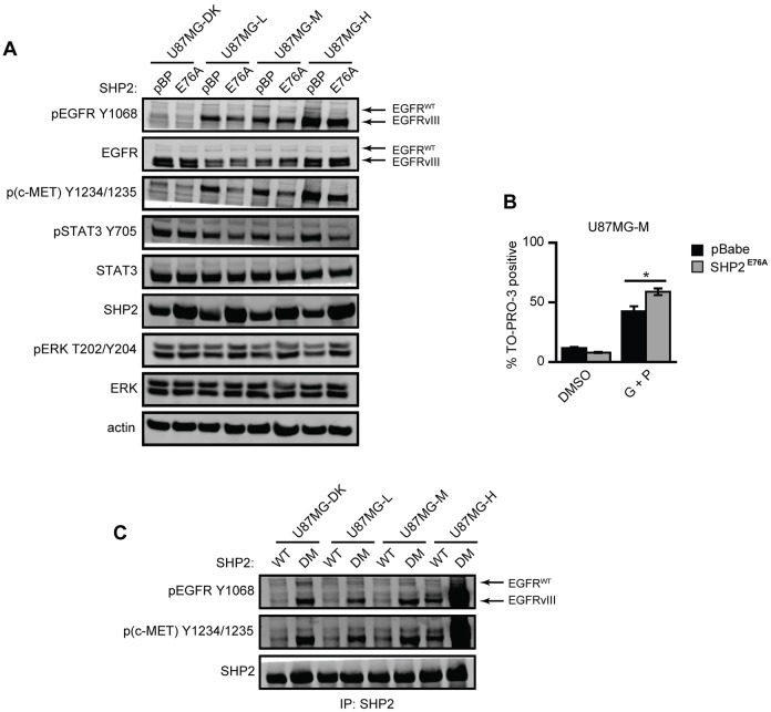
Expression of SHP2 mutants reveals negative regulation of EGFRvIII, c-MET and STAT3 phosphorylation. (A) Lysates of U87MG-DK, U87MG-L, U87MG-M and U87MG-H cells transduced with an empty pBabe.Puro vector (pBP) or SHP2E76A (E76A) and grown in full medium for 72 h were analyzed by western blotting using antibodies against the indicated proteins. Images are representative of three sets of biological replicates. (B) U87MG-M cells transduced with pBP or SHP2E76A were treated with DMSO as a control (DMSO) or with 20 µM gefitinib + 1 µM PHA665752 (G+P). After 72 h, the percentage of TO-PRO-3-positive cells was measured by flow cytometry (n = 3); *P<0.05. (C) Serum-starved cells of the indicated cell lines transiently transfected with SHP2WT or the double mutant SHP2D425A/C459S (SHP2DM) were lysed. SHP2 immunoprecipitates were analyzed by western blotting using antibodies against the indicated proteins. Images are representative of three sets of biological replicates.

