Figure 4.

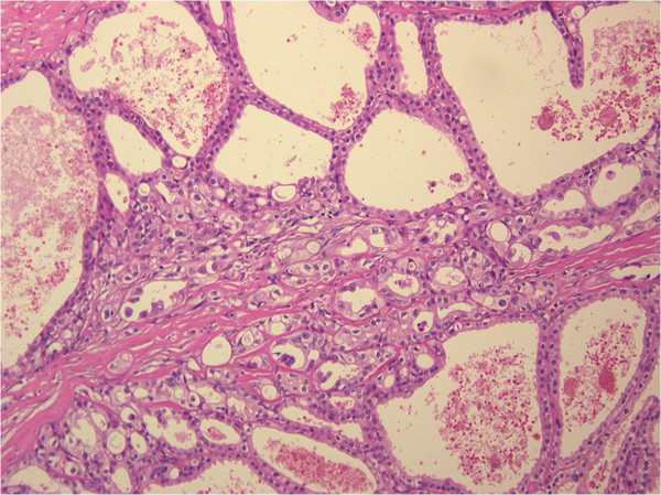
Hematoxylin-eosin staining. Hematoxylin-eosin-stained section showing cystically dilated kidney tubules lined with cuboidal and flat cells (magnification, 100×).

Hematoxylin-eosin staining. Hematoxylin-eosin-stained section showing cystically dilated kidney tubules lined with cuboidal and flat cells (magnification, 100×).