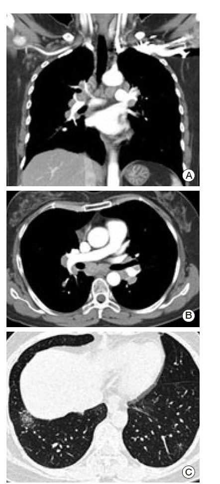
Fig. 2.

Chest computed tomography scan. (A, B) Multiple lymphadenopathies were seen on both sagittal (A) and axial (B) sections in a mediastinal setting. (C) Faint ground-glass opacities and fine reticular densities were observed on both lower lungs. These findings were also observed on both upper subpleural lungs.
