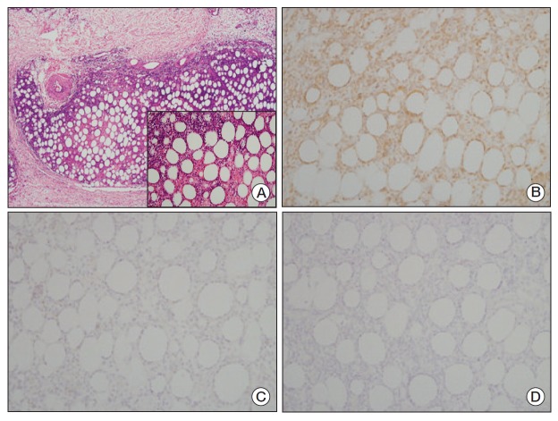
Fig. 2.
(A) The skin tissue showed diffuse atypical lymphocytic infiltration in the subcutaneous tissue similar to lobular panniculitis. Infiltration to the subcutaneous tissue without involvement of the overlying dermis or epidermis was confirmed (H&E staining, ×40). (A, inlet) The infiltrated lymphocytes had irregular, hyperchromic nuclei and indistinct nucleoli. The atypical lymphocytes were rimmed individual fat cells and some of them showed nuclear karyorrhexis (H&E staining, ×200). (B) Immunohistochemical stain, the atypical lymphocytes were positive for CD3 (×200). (C) CD20 (×200). (D) CD56 (×200). The atypical lymphocytes were negative for both CD20 and CD56.

