
Figure 1.
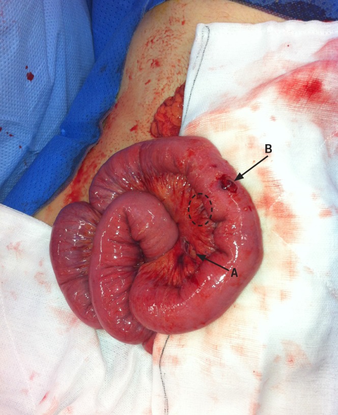
Figure 1 A partial, full thickness tear of the mesentery (A) and 10mm serosal tear on the antimesenteric border of the midileum (B). The dotted oval is the position where the third injury was later identified. (See Fig 2.)
Official websites use .gov
A
.gov website belongs to an official
government organization in the United States.
Secure .gov websites use HTTPS
A lock (
) or https:// means you've safely
connected to the .gov website. Share sensitive
information only on official, secure websites.
Figure 1 A partial, full thickness tear of the mesentery (A) and 10mm serosal tear on the antimesenteric border of the midileum (B). The dotted oval is the position where the third injury was later identified. (See Fig 2.)