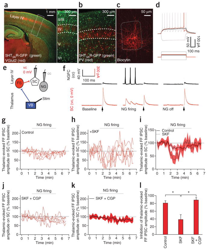
Figure 1.
Layer IV NGFC activity inhibits the canonical thalamic-evoked FFI via GABAB receptor activation. (a) Fluorescence image of a thalamocortical slice (left) of a mouse expressing 5HT3AR-GFP (layer IV delineated with VGlut2 immunostaining). Magnification (right) of the boxed area. (b) Image of 5HT3AR-GFP+ cells, stained for PV. Dashed region indicates a single layer IV barrel. (c) Dense axonal arborization of a biocytin-filled layer IV 5HT3AR-GFP+ cell. (d) Depolarizing ramp (black) and late spiking (red) of 5HT3AR-GFP+ cell at subthreshold and rheobase current injections, respectively. Gray depicts membrane responses to hyperpolarizing and 2× rheobase current injections. (e) Dual NGFC and stellate cell (SC) whole-cell recording configuration. cc, current clamp; FS, fast-spiking interneuron; NG, neurogliaform cell; VB, ventrobasal thalamus; vc, voltage clamp. (f) Single-trace examples of simultaneous current-clamp and voltage-clamp recording in an NGFC (black) and an SC (holding potential (Vh) = 0 mV; red), respectively. Left, middle and right traces are traces at baseline, during NGFC firing and after NGFC firing was turned off, respectively. Arrows denote thalamic stimulation. (g,h) Individual data showing feed-forward (FF) IPSC amplitude on SCs in control conditions (n = 5; g) and in presence of GAT-1 block by 25 μM SKF-89976A (n = 5; h). (i) Pooled data of experiments depicted in g and h. (j,k) Individual (j) and pooled (k) data showing feed-forward IPSC amplitude on SCs in presence of GAT-1 uptake block (25 μM SFK-89976A) and GABAB receptor antagonism (10 μM CGP55845A; n = 5). (l) Summary graph of reduction in FFI amplitude 1 min after onset of NGFC firing (n = 5, P < 0.05). Shaded areas in i,k and error bars in l denote s.e.m.

